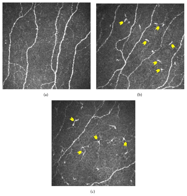
Figure 1.
Dendritic cells at the level of subbasal nerve plexus in the cornea. Subbasal nerve plexus without dendritic cells (DCs) in a healthy control eye (a). DCs without dendritic process (b) and with dendritic processes (c) in dry eye patients. Panels shown are representative IVCM images with frame size 400 × 400 microns at a depth of 45 microns.

