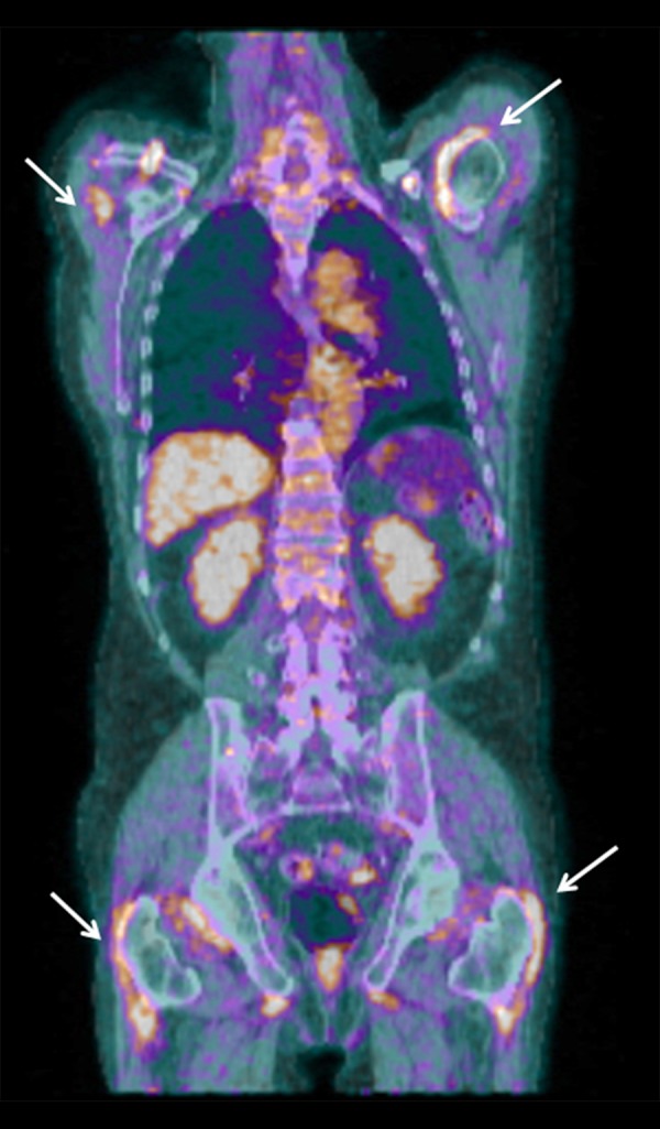
Figure 1.

Fused coronal FDG-PET/CT scan of the patient suspected of having malignancy and RS3PE syndrome. Diffusely increased FDG uptake in soft tissue around the shoulder and hip girdles (white arrows) and FDG-positive axillary lymph nodes (not shown) were suggestive of polymyalgia rheumatica. Physiologic FDG uptake can be seen in the liver and the urinary tract, but there were no other pathologic findings (i.e., no evidence of bone metastases and no lesions suspicious of malignancy). The scan was performed according to the Department of Nuclear Medicine’s standard procedure, which follows guidelines from the European Association of Nuclear Medicine. CT was performed as a low-dose scan without contrast enhancement.
