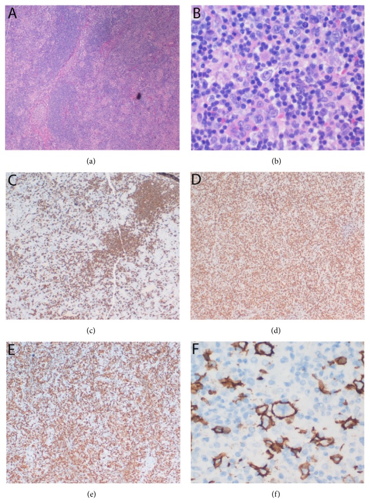
Figure 3.
THRLBCL in the right cervical lymph node. (a) The lymph node structure was almost entirely effaced by diffuse small lymphocytes with some residual follicles present (H&E, 4x). (b) Large atypical cells were surrounded by small lymphocytes and histiocytes (H&E, 40x). (c) CD20 immunostain showed the residual follicles (4x). (d) The diffuse small lymphocytes were CD3+ T lymphocytes (4x). (e) Many histiocytes were stained by CD163 (4x). (f) Immunohistochemistry for CD20 highlighted large B lymphoma cells (40x).

