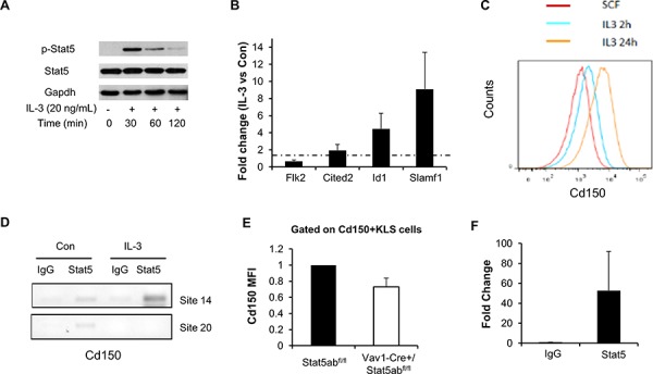
Figure 3. Stat5 activation directly regulates the myeloid differentiation marker Slamf1 in EML C1 and primary KLS cells.

A. EML C1 cells were switched from maintenance culture media (SCF alone) to differentiation culture media (SCF + IL-3) and phosphorylation of Stat5 was determined by Western blotting in cells grown under both conditions. B. Real-time PCR analysis of EML C1 cells 2 h after in vitro IL-3 exposure. Data presented (averages of four separate experiments) are fold changes of genes in IL-3 treated vs. SCF alone after the normalization to GAPDH control using ΔΔCt method. Error bars represent standard deviation. C. Staining for Cd150 in EML C1 cells after 2 h and 24 h in vitro IL-3 exposure. Cells were gated on Cd150+KLS cells. D. Stat5 was immunoprecipitated from formaldehyde-cross-linked lysates prepared from EML C1 cells treated with or without IL-3 for 2 h. Binding of Stat5 to putative Stat5 consensus binding sites 14 and 20 (control) in the Slamf1 gene was determined by PCR amplification of immunoprecipitated DNA. Nonspecific binding was assayed by rIgG isotype-control immunoprecipitation of cross-linked lysates. Results are representative of 2 independent experiments. E. Mean fluorescence intensity (MFI) of Cd150 in Cd150+KLS of Vav1-Cre/+Stat5abfl/fl and Stat5abfl/fl mice. Results are the average from four independent experiments and were normalized to Cd150 MFI for Stat5abfl/fl mice. F. Stat5 was immunoprecipitated from formaldehyde-cross-linked lysates prepared from about 1 million sorted wild type mice KLS cells. Binding of Stat5 to putative Stat5 consensus binding site 14 in the Slamf1 gene was determined by real time PCR amplification of immunoprecipitated DNA (n = 3, p = 0.08).
