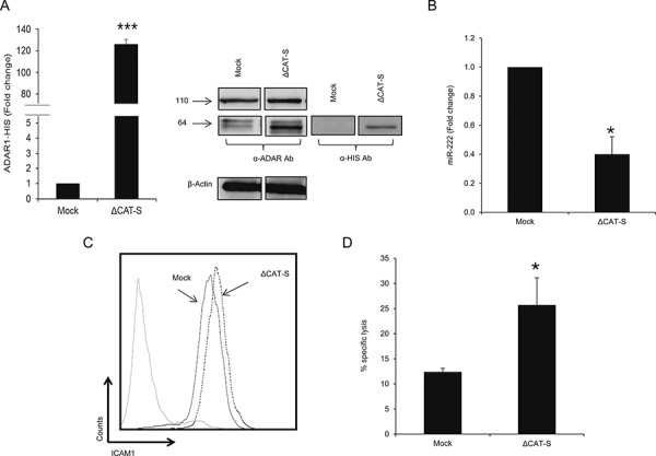
Figure 6. ADAR1 mediated-regulation is editing independent.

A. ADAR1 His-tagged mRNA levels were assessed by qRT-PCR and normalized to GAPDH expression. Protein expression of ADAR1 His-tagged constructs was determined by WB using ADAR1 polyclonal antibody and polyHistidine monoclonal antibody (cut from different regions in the same membrane). B. Expression levels of hsa-miR-222 in ΔCAT-S and Mock cells were assessed by qRT-PCR and normalized to U6 expression. C. ICAM1 protein expression in ΔCAT-S (dotted line) and Mock cells (black line) was analyzed by extracellular flow cytometry staining. D. ΔCAT-S and Mock cells were co-incubated with JKF6 for 5 h (E:T - 15:1). Specific lysis of melanoma cells was assessed by flow cytometry. Experiments were performed three times in triplicates. WB and flow cytometry figures show one representative experiment.
