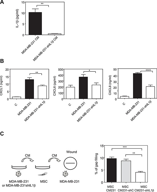
Figure 5. Inhibition of IL-1β production by MDA-MB-231 cells reduces the production of chemokines by MSCs in the presence of MDA-MB-231 conditioned medium.

A. MDA-MB-231 cells were stably transfected with empty PLKO1 vector (MDA-MB-231) or a construct against IL-1β (MDA-MB-231-shIL1β). The secretion of IL-1β MDA-MB-231 or MDA-MB-231-shIL1β was measured by ELISA. The graphs correspond to the mean ± SEM of 3 experiments. B. MSC cells were treated for 24 h with control non conditioned medium (C) conditioned medium from MDA-MB-231 transfected with empty PLKO-1 vector (MDA-MB-231) or MDA-MB-231-shIL1β cancer cells. The medium was then replaced with fresh one and collected after 24 h for ELISA assay. The levels of CXCL1, CXCL6 and CXCL8 in MSCs were measured by ELISA. Results represent the mean ± SEM of 3 independent experiments. C. The medium collected from MSCs treated with the conditioned medium control MDA-MB-231 (MSC CM231) or with MDA-MB-231transfected with sh-scramble (MSC CM231 - shC) or with MDA-MB-231-shIL1β (MSC CM231- shIL1β) in experiment B was used to treat overnight MDA-MB-231 cells. The next day, a wound was created in each well and the motility of MDA-MB-231 cells was measured by wound healing after 6 h. Left panel represents the scheme of the experiment. Results are expressed as % of gap filling and represent the mean ± SEM of 3 independent experiments.
