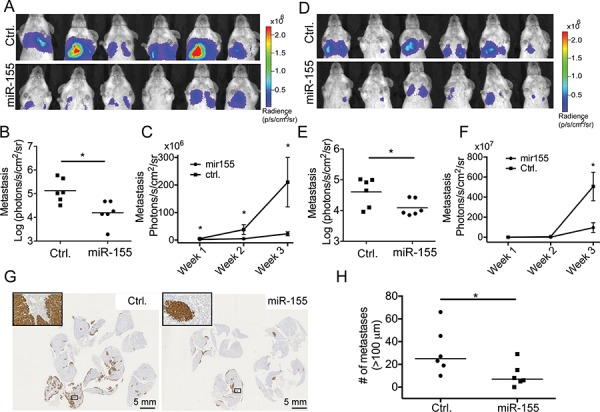
Figure 2. miR-155 decreases the tumor burden in lungs after i.v. injection.

CL16-miR-155 or CL16-Ctrl cells (7.8 × 10⁵) were injected into the tail vein of groups of female CB-17 SCID mice and tumor burden in lungs was monitored by bioluminescence imaging using an IVIS Spectrum instrument. A. IVIS images of the animals (photon radiance per area from the lung region) from the initial study (n = 6 in each group) at week 3, and B. comparison of the two groups using the Mann-Whitney statistical test (p = 0.0087). C. Increased tumor burden in lungs over time in the initial experiment measured by bioluminescence imaging starting one week after tumor cell injection. D, E. For the second animal study (n = 6 in each group), the IVIS scans (photon radiance per area from the lung region) at week 3 were also compared using the Mann-Whitney test (p = 0.041). F. Tumor growth in lungs in the second experiment measured by bioluminescence imaging over time starting one week after tumor cell injection. G. The difference in lung tumor burdens between the two groups of animals in the initial experiment was also visualized by staining the excised and FFPE lungs using an anti-human vimentin antibody. H. Quantitative evaluation of the differences in pulmonary foci/nodules (n = 6) in the initial experiment. Only tumors larger than 100 μm were included. (*p < 0.05).
