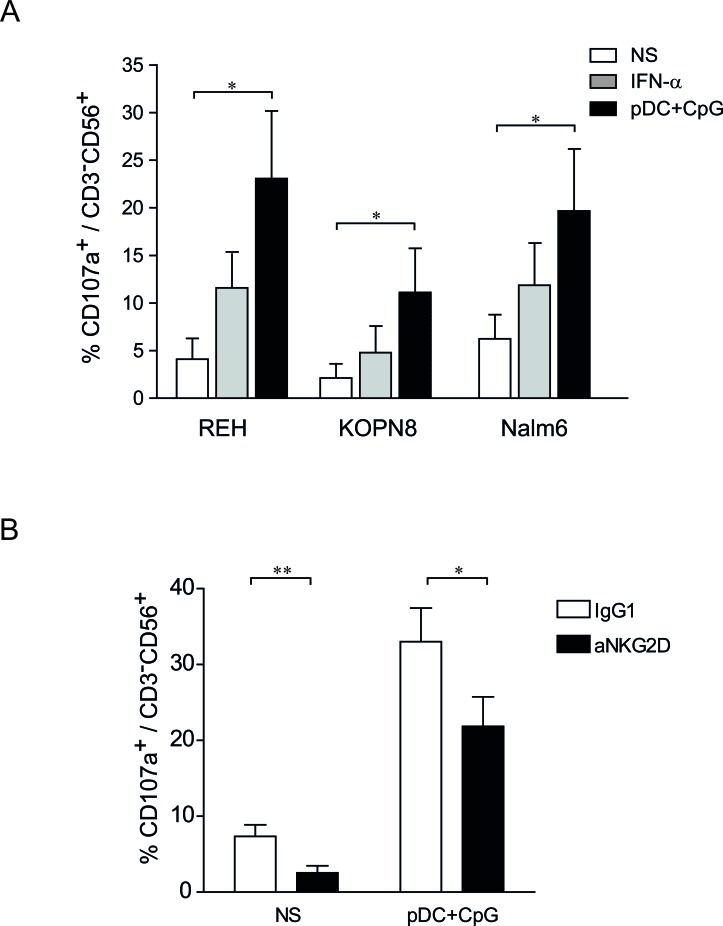
Figure 4. NK degranulation against ALL is increased following stimulation by TLR9-activated pDCs.
A. Purified human NK cells were stimulated overnight with IFN-α (1000 IU/mL), co-cultured with activated pDC or unstimulated. NK cells were then incubated with REH, KOPN8 and Nalm6 target cells at ratio 1:1 for 4 h and CD107a expression was assessed by flow cytometry. Graphs represent means ± SEM of the percentages of CD107a positive cells among CD56+CD3− cells (n = 3). B. Unstimulated NK cells (NS) and pDC-stimulated NK cells were incubated with a blocking anti-NKG2D antibody or a control IgG1 before degranulation assay. NK cells were co-cultured with Nalm6 cells (ratio 1:1) for 4 h and the proportion of CD107a+ was assessed by flow cytometry. Percentages of CD107+ cells among CD3−CD56+ cells are presented with SEM (means of 4 independent experiments). Paired t-test was used for statistical comparison *p ≤ 0.05, **p ≤ 0.01.

