
Figure 5. Knockdown of RPS3 inhibited tumor growth on melanoma in vivo.
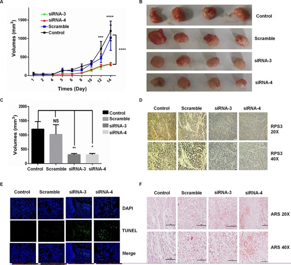
Melanoma A375 cells were injected subcutaneously into the flank of nude mice, and the visible tumors developed at the injection sites after 4 days. The DC nanoparticle-encapsulated RPS3 siRNA was then intratumorally injected twice per week. The effect of RPS3 siRNA on tumor growth curves were analyzed based on the volume at the indicated days A. At the 14th day, the tumors B. and their volumes C. were compared. The expression of RPS3 protein D. and the apoptosis in tumor tissues E. was analyzed by IHC and TUNEL assays. The ARS staining was used to analyze the concentration of Ca2+ in tumor tissues F. Data is tumor volumes ± SE in nude mice, n = 5.