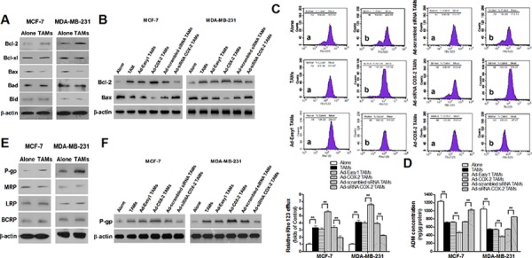
Figure 4. COX-2 in TAMs increases the expression of Bcl-2 and P-gp and decreases Bax expression in breast cancer cells.

A. The expression of Bcl-2 family members in breast cancer cells co-cultured with or without TAMs was detected by Western blot. B. The expression of Bcl-2 and Bax in breast cancer cells co-cultured with or without TAMs transfected with adenoviral COX-2 or siRNA COX-2 was detected by Western blot. C. Rho 123 efflux in breast cancer cells was analyzed by flow cytometry. The panel shows Rho 123 efflux in MDA-MB-231 cells. a, intake amount; b, residue amount. Data were expressed as a ratio of treated cells to control (Alone) cells. Mean ± SD, n = 9, **p < 0.01. D. The concentration of ADM in human breast cancer cells was measured by spectrophotometer. Mean ± SD, n = 9, **p < 0.01. E. The expression of MDR related proteins in breast cancer cells co-cultured with or without TAMs was detected by Western blot. F. The expression of P-gp in breast cancer cells co-cultured with or without TAMs transfected with adenoviral COX-2 or siRNA COX-2 was detected by Western blot. In all Western blot assays, β-actin was used as an internal loading control, and the blots shown are representative of six independent experiments.
