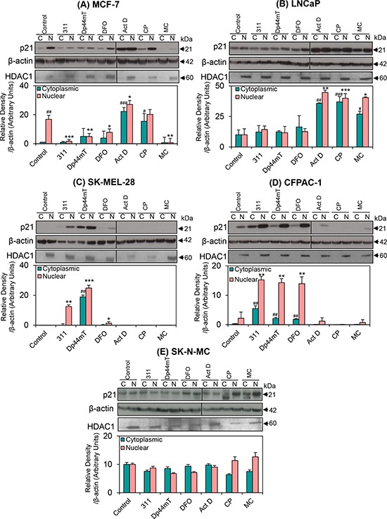
Figure 5. The effect of the chelators 311, Dp44mT, or DFO, and the DNA-damaging agents, Act D, CP or MC on the cytoplasmic and nuclear expression of p21 in five different tumor cell lines.

A. MCF-7; B. LNCaP; C. SK-MEL-28; D. CFPAC-1; and E. SK-N-MC cells. Cells were incubated for 24 h/37°C with the chelators, 311 (25 μM), Dp44mT (2.5 μM), DFO (250 μM), or the DNA-damaging agents, Act D (5 nM), CP (20 μM), or MC (30 μM). A vertical line appears on some blots to indicate the use of two gels. Indeed, in some experiments, due to hardware constraints, samples from one experiment were required to be run on 2 gels at the same time. These were then exposed equally under exactly the same conditions at the same time. The blots are typical of 3–6 independent experiments, while the densitometric analysis is mean ± SD (3–6 experiments). Relative to untreated control (cytoplasmic fraction): #p < 0.05, ##p < 0.01, ###p < 0.001. Relative to untreated control (nuclear fraction): *p < 0.05, **p < 0.01, ***p < 0.001.
