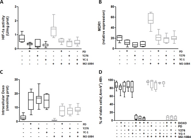
Figure 7. Specific inhibitors of RhoA, ERK1–2 and HIF-1α effectively reverse the MDR phenotype of IGHV UM cells.

IGHV UM cells were left untreated or treated with the ERK1–2 kinase inhibitor PD (10 μM), the RhoA kinase inhibitor Y276 (10 μM) and the HIF-1α inhibitor YC-1 (10 μM), both in the absence and in the presence of M2–10B4 SCs. A. HIF-1α activity. Co-culture with M2–10B4 SCs significantly increased the activity of HIF-1α (p = 0.0014). PD, Y276 and YC-1 significantly reduced the activity of HIF-1α in IGHV UM cells after 24 hours of culture, both in the absence (p always ≤ 0.0048) and in the presence (p always ≤ 0.0052) of SCs. B. MDR1 expression. Twenty four-hour co-culture with M2–10B4 SCs significantly increased the expression of MDR1 in IGHV UM cells (p < 0, 0001). After PD, Y276 and YC-1 treatment a significant decrease in MDR1 expression was observed in IGHV UM cells cultured alone (p always ≤ 0.001) and in the presence of SCs (p always ≤ 0.0001). C. Doxo intracellular accumulation. Intracellular Doxo was significantly lower in IGHV UM cells cultured with SCs than in IGHV UM cells cultured alone (p = 0.02). After 48-hour exposure to PD, Y276 and YC-1, Doxo accumulation was significantly increased, both in the absence (p always ≤ 0.003) and in the presence of M2–10B4 SCs (p always ≤ 0.0012). D. Percentage of CD19+/CD5+ viable cells. IGHV UM cells were exposed to 1 μM Doxo, used alone and in combination with each inhibitor (i.e. PD+Doxo, Y276+Doxo, YC-1+Doxo), both in the absence and in the presence of the M2–10B4 SCs. Cell viability was determined by Ann-V staining and cytofluorimetric analysis on CD19+/CD5+ cells after 48-hours of culture. Doxo alone, as well as the three inhibitors used as single agents, did not induce a decrease in cell viability. By contrast, the combinations PD+Doxo, Y276+Doxo and YC-1+Doxo significantly reduced the viability of IGHV UM cells both in the absence and in the presence (p always < 0.0001) of SCs. In panels A–D results are from 7 side-by-side experiments. Box and whiskers plot represent median values, first and third quartiles, and minimum and maximum values for each dataset.
