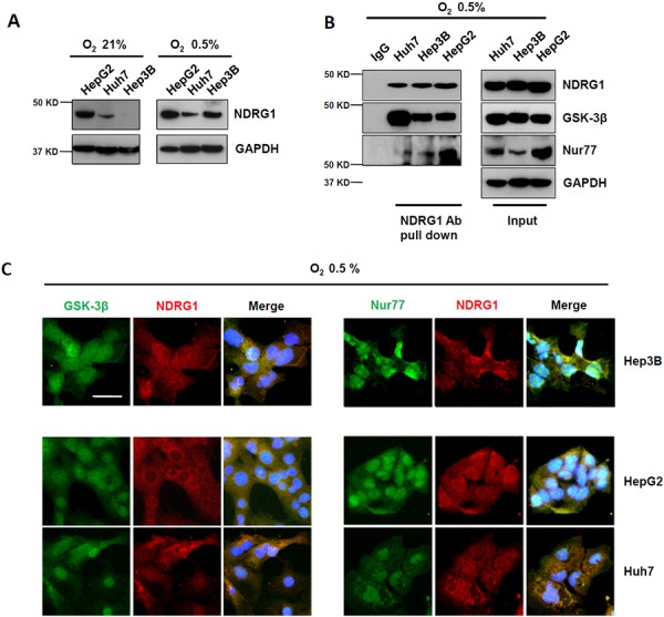
Figure 2. Hypoxia-inducible NDRG1 expression and its interactions with GSK-3β and Nur77 in HCC cells.

A. Western blot showing that NDRG1 expression was increased under hypoxia (0.5% O2) in HCC cells. GAPDH was used as loading control. B. Co-IP showing interaction of NDRG1 with GSK-3β and with Nur77 in HCC cells under hypoxia. C. Immunofluorescence staining showed the co-localization of NDRG1 and GSK-3β or Nur77 in HCC cells under hypoxia (400× magnification; scale bars: 5 μm).
