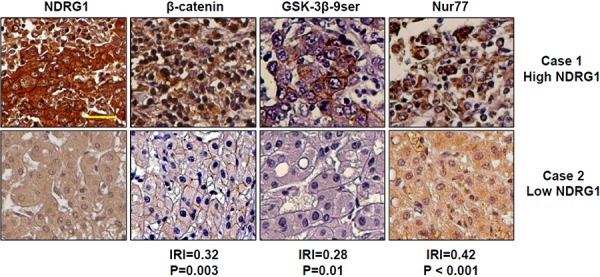
Figure 7. Expression of NDRG1, β-catenin, GSK-3β 9ser, and Nur77 in HCC patients.

Immunohistochemistry staining of NDRG1, β-catenin, GSK-3β 9ser and Nur77 were performed in 82 cases of HCC represented on tissue microarrays. Representative images of two HCC patients (Case 1 with high NDRG1, and Case 2 with low NDRG1) are shown for each protein. Based on semi-quantitative signal intensities (0-negative; 1- low expression, positive cells present in < 50% of entire area; 2- high expression, positive cells present in > 50% of entire area); correlation plots of NDRG1 with each protein shows that NDRG1 protein expression is positively correlated with β-catenin (│R│= 0.32, p = 0.003), GSK-3β 9ser (│R│= 0.28, p = 0.01), and Nur77 (│R│= 0.42, p < 0.001) (200× magnification; scale bars: 10 μm).
