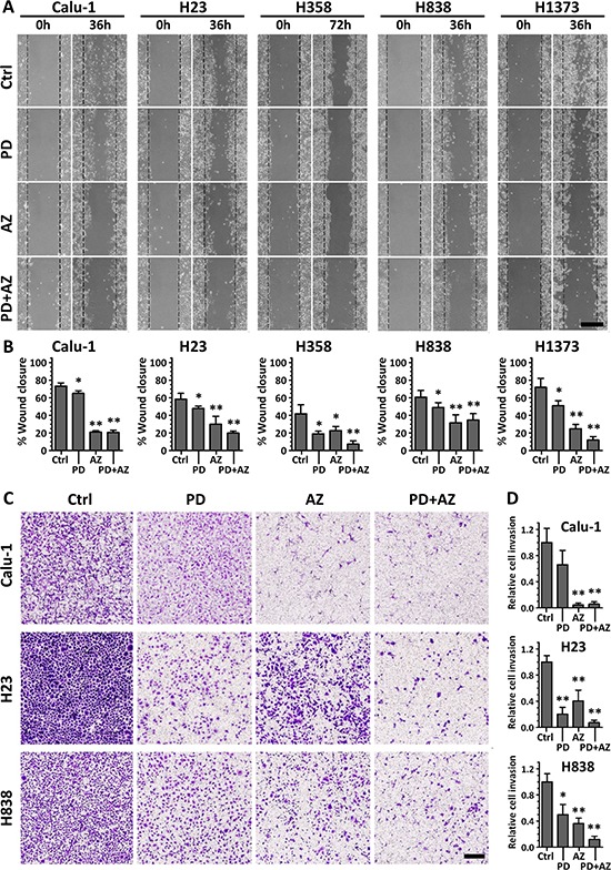
Figure 4. PD0325901 and Saracatinib and the combination treatment reduced cell migratory and invasive activity.

Cell lines were treated with DMSO control (Ctrl), or with PD (1 μM), AZ (4 μM) or their combination. A. for cell migration assays, the wounded areas were imaged at 0 h and 36 or 72 h after the monolayer cultures were scratched. Scale bar: 500 μm. B. the gap area of each image was measured, and the wound closure percentages (mean ± SD) were calculated from at least three independent experiments. C. for Boyden chamber Matrigel invasion assays, cells that invaded through to the underside of the transwell filter after 48 h incubation under various drug treated conditions were fixed, crystal violet stained and imaged. Scale bar: 250 μm. D. cell count analysis was performed for each image and the relative cell invasion (mean ± SD) of each treated group compared to the Ctrl group of each cell line was calculated from at least three independent experiments. *,P < 0.05; **,P < 0.01 compared with Ctrl group.
