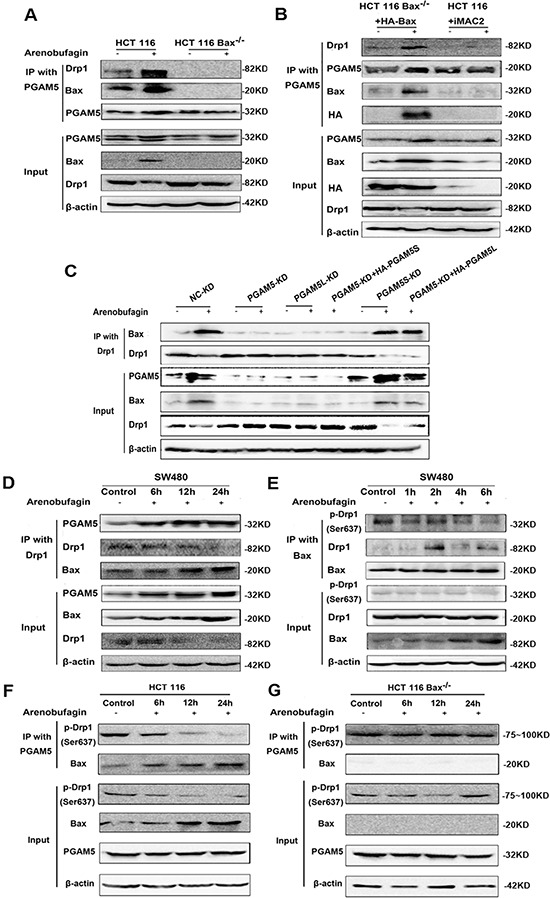
Figure 6. Bax Interacts with PGAM5-Drp1 Complex.

A. HCT116 WT and HCT116 Bax−/− cells were treated with 1 μM arenobufagin for 12 h. IP was performed with an anti-PGAM5 antibody. Co-IP Drp1 and Bax were detected by western blotting (n = 3). B. HCT116 Bax−/− cells were transfected with the control vector or HA-Bax, and treated with 1 μM arenobufagin for 12 h. HCT116 WT Cells treated with the combination of arenobufagin and iMAC2 (20 μM). Cells were lysed and immunoprecipitated using an anti-PGAM5 antibody. Immunoprecipitates were subjected to western blotting using Drp1 and Bax antibody (n = 3). C. PGAM5-KD HCT116 WT cells were transfected with PGAM5L or PGAM5S coding sequence, and the cells were then treated with arenobufagin for 12 h. IP was performed with a PGAM5 or Drp1 antibody. Co-IP PGAM5, Drp1 and Bax was detected through western blotting (n = 3). D. SW480 cells were treated with 1 μM arenobufagin for different time, and then IP was performed with Drp1 antibody; Co-IP PGAM5, Drp1 and Bax was detected by western blotting (n = 3). E. SW480 cells were treated with 1 μM arenobufagin for different time, and then IP was performed with Bax antibody; Co-IP p- Drp1 (ser 637), Drp1 and Bax was detected by western blotting (n = 3). F–G. HCT116 WT and HCT116 Bax−/− cells were treated with 1 μM arenobufagin for different time. IP was performed with an anti-PGAM5 antibody. Co-IP p- Drp1 (ser 637) was detected by western blotting (n = 3).
