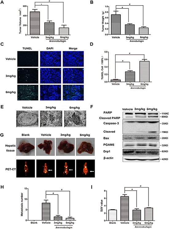
Figure 8. Arenobufagin inhibits growth and metastasis of orthotopically implanted colorectal carcinoma through inducing apoptosis.

A. Isolated tumor size and B. Tumor weight from the SW480-eGFP mouse Orthotopic CRC model. C–D. Tumors were excised and processed for immunostaining with TUNEL Kit(green) and DAPI (blue), and fluorescent images were obtained by microscopy, 400× for all, scale bar = 150 μm. E. Morphological changes of arenobufagin-treated tumors as observed by TEM. 15000× for all, scale bar = 2 μm. F. PARP, Caspase-3, Drp1, PGAM5 and Bax in tumor tissue lysates from vehicle- and arenobufagin-treated mice were detected by western blot analysis. G. The image of metastasis in mice hepatic tissue (above) and PET-CT images (below). H. Bar graphs at the bottom show the percentages of metastasis number in mice hepatic tissue. I. Ratio of 18F-FDG SUV of drug treatment versus control in whole body for PET-CT mice. *P < 0.05, #P < 0.01, n = 6, one-way ANOVA, post hoc comparisons, Tukey's test. Columns, means; error bars, SEs. See also Supplementary Figure S5 and Supplementary Video S1–S4.
