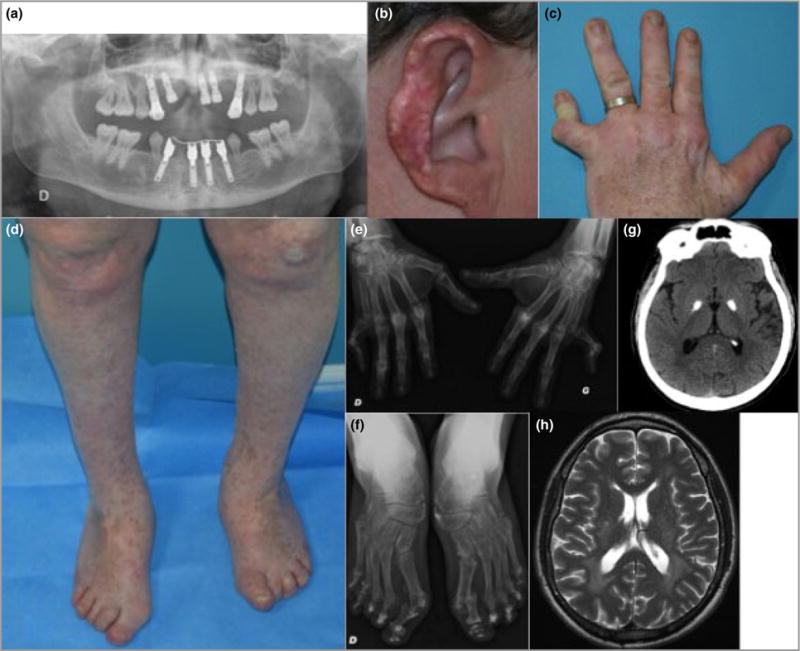
Fig 2.

Clinical images of the 41-year-old father. (a) Pantomogram showing loss of permanent teeth. The remaining dentition, molars and premolars demonstrate root hypoplasia. (b) Scarring and tissue loss of the outer ear helix following chronic ulceration. (c) Deformities of the metacarpophalangeal and interphalangeal joints of the left hand, and multiple lentigines. (d) Hallux valgus, psoriatic lesions over the knees and multiple lentigines. (e) Radiography showing joint deformities on the hands. (f) Radiography showing joint deformities on the feet. (g) Brain imaging at age 41 years showing bilateral calcification of the globus pallidus on computed tomography. (h) Brain imaging at age 41 years illustrating high signal in the white matter around the posterior poles of the lateral ventricles on axial T2-weighted magnetic resonance imaging.
