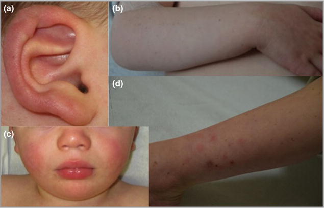
Fig 3.

Clinical images of the younger sibling of the proband. (a) Erythema and ulceration of the outer helix of the right ear. (b) Multiple lentigines of the right forearm. (c) Bilateral erythema of the cheeks. (d) Lower-limb atrophic scars, which occurred following resolution of papular lesions.
