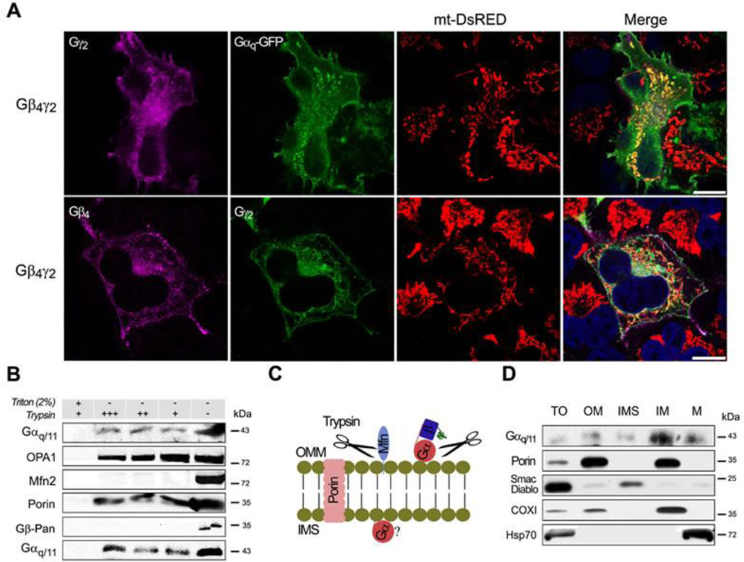
Fig. 2. Gαq and different Gβγ dimers localized at the mitochondria.
(A) Confocal micrographs of HeLa cells stably expressing mt-DsRed and Gβ4-Flag γ2-HA and/or Gαq, immunostained with anti-Flag or HA and mounted with DAPI. (B) Mitochondrial fractions of NIH3T3 cells submitted to trypsin digestion in presence or absence of Triton X-100, immunoblotted with anti-Gαq/11, OPA1 (inner membrane), Mfn2 (outer membrane), Porin (integral outer membrane) and Gβ-Pan (representing Gβγ dimer). (C) Diagram showing the likely actions of trypsin on proteins blotted in B. (D) Mouse liver mitochondrial sub-fractions shown the presence of Gαq/11 at the inner membrane, immunoblotted with Gαq/11 and markers: Porin (OM), Smac/Diablo (IMS), COXI (IM) and Hsp70 (M).

