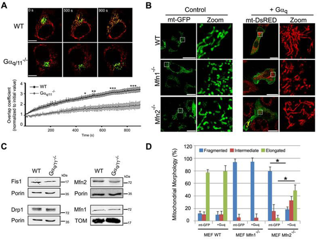
Fig. 4. Impairment in mitochondria fusion in absence of Gαq/11.
(A) MEFs transfected with mt-DsRed and mito-PAGFP. Mito-PAGFP was photoactivated, mt-DsRed was photobleached at t=0 s. Panels show the same cell at a range of time points. Scale bar: 75 µm. Data show mean ± s.e.m (n=5). ANOVA (*p<0.05, **p<0.01 and ***p<0.001) was employed. (B) Confocal micrographs of MEFs transfected with mt-GFP or Gαq/mt-DsRED, immunostained with anti-Gαq/11 antibody (right panel in green). Zoom shows only mitochondria. Scale bar: 25 µm. (C) Mitochondrial morphology was scored from B. Data represent mean ± s.d. (n=50) of three independent experiments. Chi-Square test was employed (*p<0.0001). (D) Confocal micrographs of MEFs in presence of Gβ2-Flag (green on left and purple on right panel)/mt-dsRED with or without Gαq-GFP (green on right panel) mounted with DAPI (blue). Zoom shows only mitochondria.

