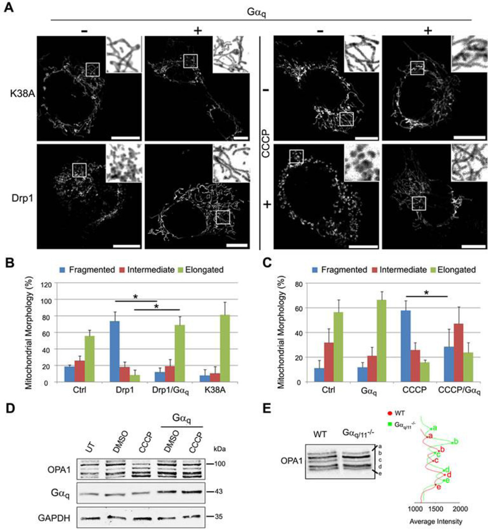
Fig. 5. Gαq stabilizes mitochondrial fusion, blocking fragmentation induced by Drp1 or CCCP.
(A) Confocal micrographs of MEF wild-type cells transfected with mt-DsRed (grey) and Drp1-HA or Drp1-(K38A)-HA or treated with 10 µM CCCP (+) or DMSO (−) for 3h, overexpressing (+) or not (−) Gαq, immunostained with anti-HA and anti-Gαq/11 (not shown). (B-C) Mitochondrial morphology quantified as mentioned in Figure 4C. Chi-Square test was employed (*p<0.0001). Experiments were carried out as in A. Data represent mean ± s.d. (n=50) of three independent experiments. (D) MEF cells were transfected with pcDNA3 or pcDNA3-Gαq and the day after incubated for 3h with 10 µM CCCP or DMSO. Lysates were immunoblotted with the indicated antibodies. (E) MEF WT and Gαq/11−/− cells immunoprecipitated for OPA1 isoforms and immunoblotted with OPA1 antibody, quantified by Line Profile of Odyssey System.

