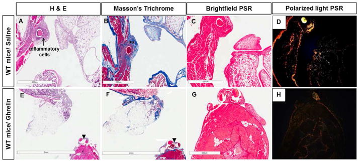
Figure 4. Ghrelin reduces inflammatory infiltrate and fibrotic thickening in peritoneal ischemic buttons.
Photomicrographs of surgical adhesion model of saline (A, B, C, D) and ghrelin-treated wild type mice (E, F, G, H). H&E (A, E), Masson’s trichrome (B, F) and Picrosirius red (PSR) (C, D, G, H) staining. Saline-treated mice showed peritoneal wall thickness with thickened collagen bundles (**) and infiltration of inflammatory cells (arrow) around the suture (A, B). In comparison, ghrelin-treated animals showed reduction of fibrotic thickening and collagen deposition around the sutures (arrow heads) (F). Photomicrographs of Picrosirius red-stained sections with polarized light used to distinguish collagen type I (yellow and orange fibers) from Collagen type III (green fibers) (D, H). The amount of collagen I deposition after surgery was strongly higher in the saline-treated samples (D) than in the ghrelin-treated ones (H). Bars, 600 μm (A, C, D, H), 500 μm (B, G) and 2 mm (E, F).

