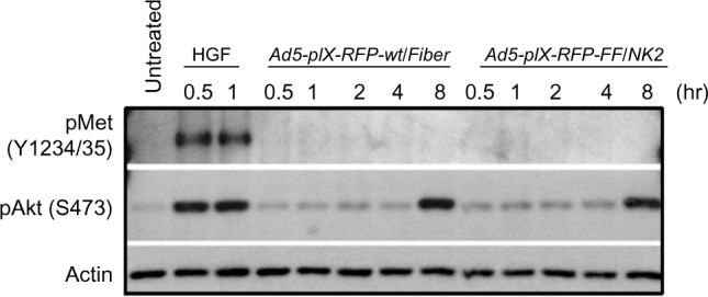
Figure 9.

Effect of Ad5-pIX-RFP-FF/NK2 on cMet autophosphorylation.
Notes: The Hep3B2.1 cells were infected with either Ad5-pIX-RFP-FF/NK2 or Ad5-pIX-RFP-wt/Fiber at an MOI of 500 VP/cell. Cells treated with HGF (33 ng/mL) were used as a positive control. Cell lysates at each time point were separated on 10% SDS-PAGE gels, and the proteins were then transferred onto PVDF membranes, blocked, and probed with a monoclonal antibody against phospho-cMet. An anti-β-actin antibody was used as a loading control.
Abbreviations: MOI, multiplicity of infection; VP, viral particles; HGF, hepatocyte growth factor; SDS-PAGE, sodium dodecyl sulfate-polyacrylamide gel electrophoresis; PVDF, polyvinylidene fluoride; hr, hour; Ad5, adenovirus serotype 5; NK2, a secreted truncated splicing variant that extends through the second kringle domain.
