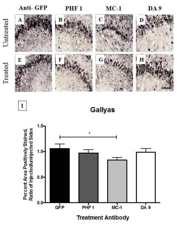
Fig. 1. Gallyas silver staining of hippocampus, four days after intracranial injection of anti-tau antibodies into rTg4510 mice.
One-year old rTg4510 mice (n = 6 – 8 animals per group) received intracranial injection (by convection enhanced delivery, CED) of potential tau-treatment antibodies into the right hemisphere. The left hemisphere remained untreated. Three different types of antibodies were tested, in comparison to an anti-GFP control injection. PHF-1 is a phosphorylated form of tau, MC-1 is a conformation-dependent antibody, and DA-9 is a pan-tau antibody. MC-1 treatment demonstrated a reduction in NFTs (p < 0.05 by ANOVA), while the other antibodies did not. Scale bar = 100 μm

