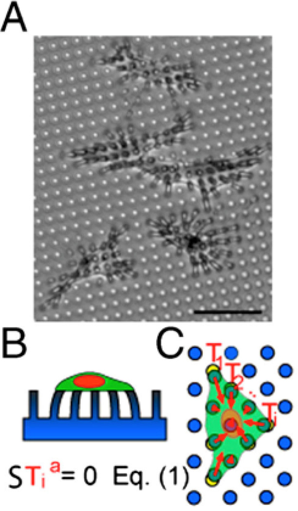
Figure 3. Micropost arrays.
A) Endothelial cells tug on microposts (dia. = 3 µm). The microposts were coated with fibronectin by microcontact printing to restrict cell adhesion to a specific area. Scale bar is 50 µm. B) Side view cartoon of the cell on the micropost array. C) The individual traction force vectors exerted by the cell sum to zero. Reproduced with permission from [200].

