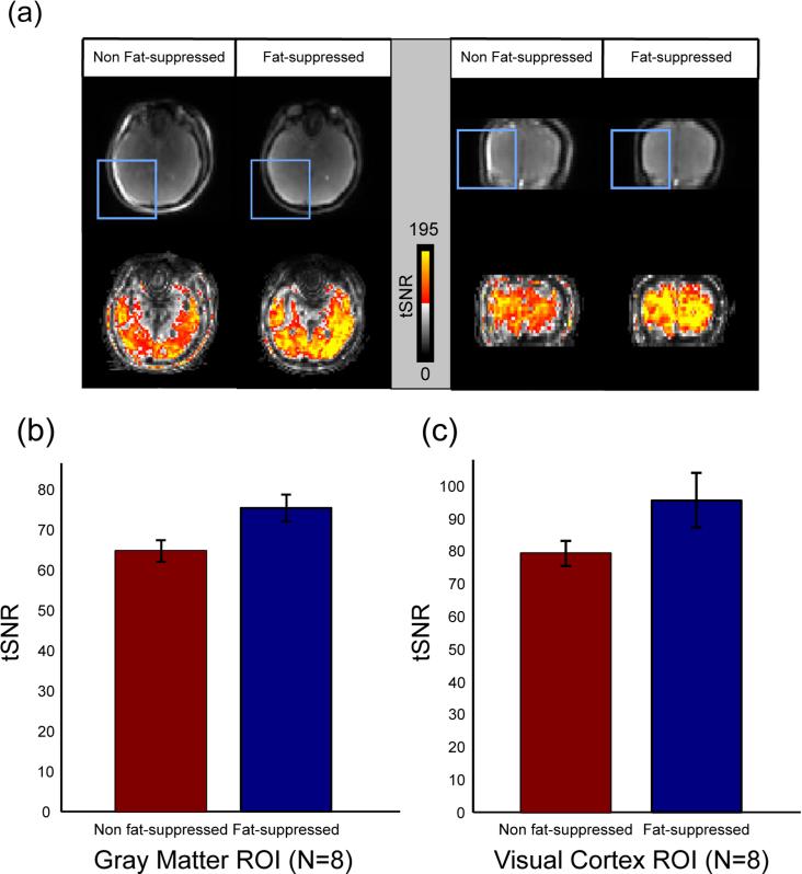
FIG. 6.
Observed temporal SNR (tSNR) increase in fat-suppressed alt-SSFP results compared to non fat-suppressed results. a: Select slices from a breath-holding study of a representative subject. Raw images (top) show bright fat signal shift into the brain volume (in boxes) without fat suppression (left: axial, right: coronal). By using the designed short SPSP pulse, the bright fat signal is well suppressed. tSNR maps show that applying fat suppression increases temporal stability in fat-shift artifact regions and overall. b: The mean tSNR was calculated for all subjects over gray matter mask ROI over N = 8 subjects. Fat-suppressed results have an increase of 17% ± 4% mean tSNR over non fat-suppressed results. c: Mean tSNR was computed within the V1 region of the visual cortex over N = 8 subjects. Fat-suppressed results have an increase of 20% ± 8% over non fat-suppressed results.

