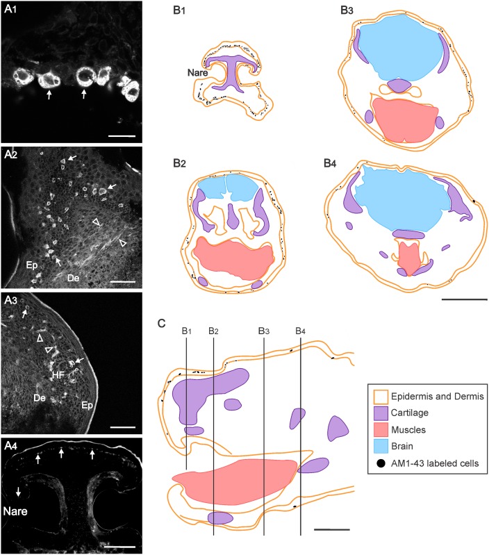
Fig 3. Distribution of AM1-43 labeled cells in the head of newborn opossums.
(A) Microphotographs of head sections showing AM1-43 labeled cells (arrows) and nerve fibers (empty arrowheads) (A1-A4). Microphotographs in panels A1-A3 were obtained with a laser scanning confocal microscope (see Materials & Methods). (B) Schematic drawing of a sagittal section of the head of a newborn opossum indicating the levels at which the drawings in C are taken. (C) Cross sections from the head of another newborn opossum at the rostrocaudal levels indicated in B, illustrating the position of AM1-43 labeled cells. Legends: De, dermis; Ep, epidermis; HF, developing hair follicle. Scalebars: 20 μm in A1; 50 μm in A2 and A3; 200 μm in A4; 500 μm in B and C.

