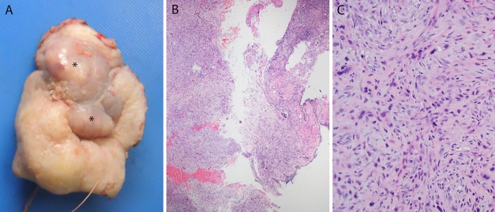
Fig. 3.
Spindle cell (sarcomatoid) carcinoma. A gross photograph of a total rhinectomy specimen for spindle cell carcinoma a shows violaceous nodules of tumor (asterisks) bulging underneath the skin of the nasal bridge. Histologically, the tumor was biphasic, consisting predominantly of keratinizing type squamous cell carcinoma but with sheet-like areas of spindle cells (b-×4 magnification) in a vaguely fascicular pattern with hyperchromatic, pleomorphic nuclei, collagenous to myxoid stroma (c-×20 magnification) and abundant mitoses, including atypical ones

