Fig. 3.

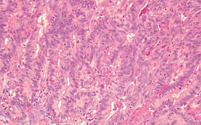
Intestinal-type adenocarcinoma, colonic growth pattern. Glandular structures and some trabecular areas. High mitotic activity. H–E stain ×250

Intestinal-type adenocarcinoma, colonic growth pattern. Glandular structures and some trabecular areas. High mitotic activity. H–E stain ×250