
Fig. 2A–D.
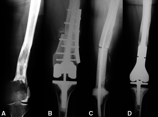
A 42-year-old female patient had a giant cell tumor of the left distal femur. She underwent reconstruction with an APC after resection of the tumor. (A) An AP radiograph of the left distal femur shows the epiphyseal tumor with fracture of the distal femur. (B) An AP radiograph obtained after resection of the tumor and reconstruction with an APC shows fixation of the allograft to the host bone with a lateral plate. (C) An AP radiograph of the left distal femur 6 months after reconstruction shows a temporary antibiotic-impregnated cement spacer that replaced the original APC owing to a deep infection. (D) Three years postoperatively, the patient’s AP radiograph of the left distal femur is shown. The patient underwent reconstruction with a modular cemented endoprosthesis.