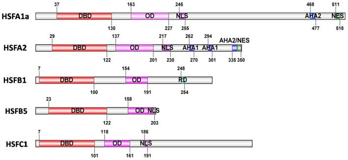
Figure 1.
Basic structure of HSFs. The block diagrams represent five tomato HSFs with their conserved functional domains. The conserved domains are identified by Heatster (http://www.cibiv.at/services/hsf/). DBD, DNA binding domain; OD, oligomerization domain (HR-A/B region); NLS, nuclear localization signal; NES, nuclear export signal; AHA, activator motifs; RD, tetrapeptide motif–LFGV–as core of repressor domain. (Adapted from Scharf et al., 2012).

