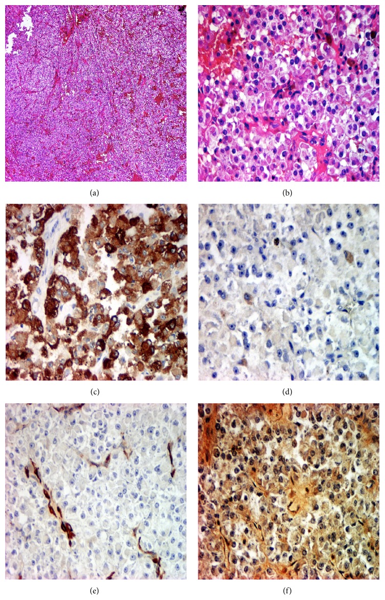
Figure 4.
Histology: (a) trabecular architecture and nested growth pattern. HE, 40x. (b) Cell proliferation with an eosinophilic or clear granular cytoplasm; a prominent vasculature composed of delicate thin-walled capillaries was also appreciable. HE, 200x. (c) Immunohistochemical staining showing positivity for HMB45. 200x. (d) Immunohistochemical staining showed negativity for S100 protein. 200x. (e) Immunohistochemical staining for α-SMA showed slight staining in tumour cells. 200x. (f) Immunohistochemical staining showed focal positivity for Calponin. 200x.

