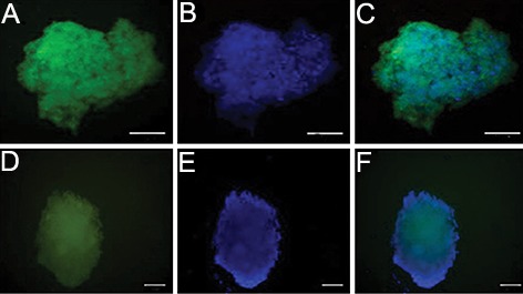
Fig.4.

Immunofluorescence reveals bone marrow derived SSEA-1 positive cells differentiated into insulin secreting cells in vitro. The insulin secreting cells at days 24 were fixed and stained with antibodies against Pdx1 and Glut2 and visualized with secondary antibody (FITC). 4′, 6-diamidino-2-phenylindole (DAPI) was used to counter-stain DNA (blue) (scale bar; 50 μm).
A. AntiPdx1 immunofluorescence, B. Nuclear counterstain with DAPI, C. Merged image of A and B, D. AntiGlut2 immunofluorescence, E. Nuclear counterstain with DAPI and F. Merged image of D and E. SSEA-1; Stage-specific embryonic antigen 1.
