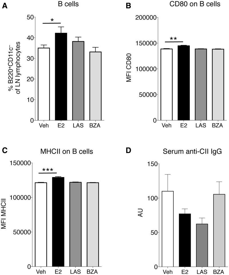
Fig. 6.
Analysis of lymph node B cells and serum CII antibodies
OVX DBA/1 mice were subjected to CIA and treated with LAS, BZA, E2 or vehicle. Lymph node cells were analysed using flow cytometry, defining (A) the percentage of B cells (B220+CD11c−), (B) MFI of CD80 on the B220+CD11c−CD80+ population and (C) MFI of MHC class II on the B220+CD11c−MHCII+ population. (D) Serum concentrations of IgG antibodies against collagen type II were assessed by ELISA. Bars represent mean (s.e.m.). Differences between treatments and vehicle were analysed using analysis of covariance with experiment day as the covariate and Dunnett’s post hoc test (A and C) or analysis of variance and Dunnett’s post hoc test (B and D). n = 13–16 mice/group. *P < 0.05, **P < 0.01, ***P < 0.001. BZA: bazedoxifene; E2: 17β-estradiol; LAS: lasofoxifene; MFI: mean fluorescence intensity; OVX: ovariectomised; veh: vehicle.

