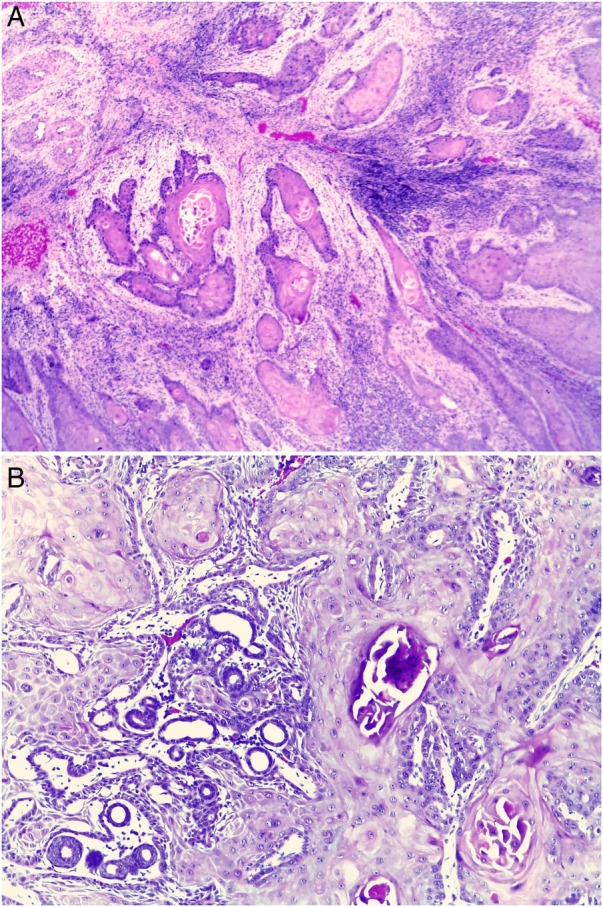
Figure 4.

(A) Microscopic view of the main tumour. Carcinomatous infiltrating component along with cytological atypia and mitosis. (H&E ×100). (B) Microscopic view of the main tumour. Carcinomatous infiltrating component, dissecting eccrine sweat glands. (H&E ×200).
