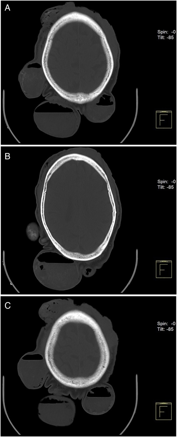
Figure 5.

(A–C) CT scan of the head. Five epicranial oval lesions of various sizes, the smallest lesion being 14.5×15 mm, and the largest, 85×80 mm in diameter, with non-pure heterogeneous liquid-air content, forming multiple fluid-air levels. There are no discernable signs of regional tumour invasion.
