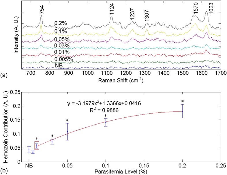
Figure 1. SERS spectra and hemozoin contribution in Method 1.
(a) SERS spectra contributed by hemozoin in infected blood treated by Method 1. (b) Hemozoin contribution as a function of parasitemia level. In (a), the data point corresponding to a parasitemia level of 0.01% is marked by a red box. In (b), the resulting curve of the second-order polynomial fitting for the data points corresponding to parasitemia levels in the range of 0.01% to 0.2% is shown in red. The data corresponding to normal blood samples, labeled as “NB”, is added manually to facilitate comparison. The y axes in both figures are plotted in arbitrary units, labeled as “A. U.” The asterisks indicate parasitemia levels at which the Raman peak intensity at 1623 cm−1 were significantly different from that in the normal blood sample in t-test (p < 0.05). The detection limit was determined to be 0.01% parasitemia level (marked by the red box) in this manner.

