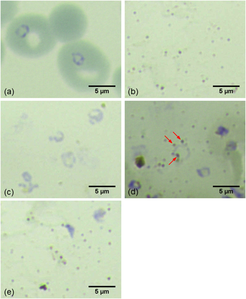
Figure 3. Giemsa stained images of blood samples treated by Method 1 and Method 2.
(a) a blood sample with parasites in the ring stage prior to cell lysis, (b) silver nanoparticles alone, (c) a blood sample after blood cell membrane lysis but prior to nanoparticle synthesis treated by Method 2, (d) the blood sample after nanoparticle synthesis treated by Method 2 and (e) a blood sample treated by Method 1 and then mixed with nanoparticles synthesized separately.

