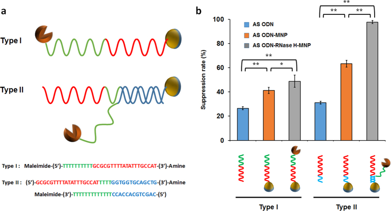
Figure 4. Design and comparison of molecular nanodevices consisting of ASODN, RNase H and magnetic nanoparticles.
(a) Schematic of the two designed MNP conjugates with ASODN and RNase H. Red letters, ASODN regions; green letters, linker regions; blue letters, complementary regions. (b) Comparison of suppression rates of the two designed nanodevices during cell-free protein synthesis of sfGFP. Final concentration of ASODN was set at 2 μM and approximately same concentrations of RNase H was conjugated on MNPs. Error bars in the graph indicate standard deviations from three independent reaction samples. *p-value < 0.05. **p-value < 0.01.

