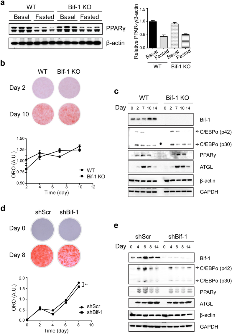
Figure 4. Bif-1 deficiency does not affect adipogenesis.
(a) Bif-1 does not impact adipogenesis in vivo. Relative protein expression levels of PPAR-γ in WT and Bif-1 KO WAT homogenates prepared from 6- week-old mice were determined via immunoblotting (n = 3). (b) Immortalized perivascular cells isolated from brown adipose tissue (iPBA) of 6-week-old mice as described in Methods were induced to differentiate with an adipogenic cocktail. Cells were subjected to Oil Red O (ORO) staining on the indicated days following adipogenesis induction. Representative images were acquired, and the optical density of ORO extracted from the cells was quantified by absorption at 520 nm (n = 4). (c) Lysates prepared from iPBA cells on the indicated days during adipogenesis were subjected to immunoblotting with the indicated antibodies. (d) 3T3-L1 cells transduced with lentiviruses encoding Bif-1 shRNA (shBif-1) or scrambled shRNA (shScr) were induced to differentiate as described in Methods Cells on indicated days during adipogenesis were stained with ORO. Representative images were acquired, and the optical density of ORO extracted from the cells was quantified by absorption at 520nm (n = 4). Statistical significance was determined using Student’s t-test. All values are mean ± SEM. Differences with controls were significant for **p < 0.01. (e) Lysates prepared from 3T3-L1 shScr and shBif-1 cells on the indicated days during adipogenesis were subjected to immunoblotting with the indicated antibodies.

