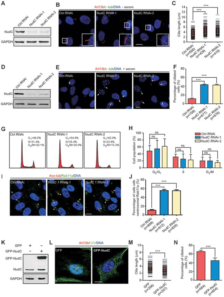
Figure 2.
NudC suppresses ciliogenesis in mammalian cells. (A-C) RPE-1 cells treated with either NudC RNAi-1 or -2 under serum starvation are subjected to the following analyses. Immunoblotting confirms significant knockdown of NudC (A). GAPDH is the internal control. Immunofluorescence with anti-Arl13b (cilia marker) and γ-tubulin (γ-tub) antibodies reveals an increase in cilia length after NudC depletion (B). Boxed regions are enlarged in insets. Lengths of more than 120 cilia (per experiment) are measured by Image J software (C). (D-J) RPE-1 cells treated with either NudC RNAi-1 or -2 are analyzed. Western blotting shows significant suppression of NudC (D). Immunostaining reveals spontaneous ciliogenesis (arrow) in NudC knockdown cells (E). Percentage of cells with cilia in each experiment (F). Flow cytometry reveals no significant difference between control and NudC-depleted cells during cell cycle progression (G, H). Immunofluorescence with anti-acetylated tubulin (ace-tub, cilia marker) and Rab11a antibodies reveals an increase in Rab11a-positive vesicles around the centrosome regions (arrowheads) in NudC-depleted cells (I, J). More than 150 cells are scored each time. (K-N) Overexpression of NudC suppresses ciliogenesis. RPE-1 cells transfected with the indicated vectors are subjected to western blotting (K) and immunostaining (L). Lengths of more than 60 cilia (per experiment) are measured by Image J software (M). Over 180 cells with cilia are counted in each experiment (N). DNA is visualized by DAPI. Scale bars represent 10 μm. Quantitative data from at least three independent experiments are shown as mean ± SD. n, sample size. ***P < 0.001; ns, not significant (P > 0.05).

