Figure 5.
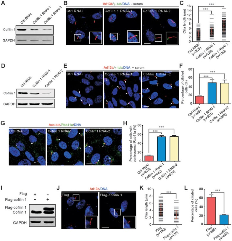
Cofilin 1 suppresses cilia assembly in mammalian cells. (A-C) RPE-1 cells were transfected with siRNA oligos, cofilin 1 RNAi-1 or -2, targeting different regions of cofilin 1 mRNA. Western blotting shows efficient knockdown of cofilin 1 (A). GAPDH is the internal control. Immunofluorescence with anti-Arl13b and γ-tubulin (γ-tub) antibodies reveals an increase in cilia length after cofilin 1 depletion (B). Insets are higher magnifications of the boxed areas. Cilia length (more than 70 cilia in each experiment) is scored using Image J software (C). (D-H) RPE-1 cells were treated with cofilin 1 RNAi. Western blotting confirms significant depletion of cofilin 1 (D). Immunostaining with antibodies reveals spontaneous ciliogenesis (arrows) in cofilin 1 knockdown cells (E). Percentage of cells with cilia in each treatment (F). Immunofluorescence reveals Rab11a-positive vesicles are frequently accumulated around the centrosome regions (arrowheads) in cofilin 1-depleted cells (G, H). More than 150 cells are scored each time. (I-L) Overexpression of cofilin 1 suppresses ciliogenesis. Western blotting (I) and immunostaining (J) of RPE-1 cells transfected with the indicated vectors. Insets show higher magnifications of cilia. Cilia length (more than 40 cilia in each experiment) is measured using Image J software (K). Percentage of cells with cilia in each treatment (L). DNA is labeled with DAPI. Scale bars represent 10 μm. Quantitative data from at least three independent experiments are shown as mean ± SD. n, sample size. ***P < 0.001.

