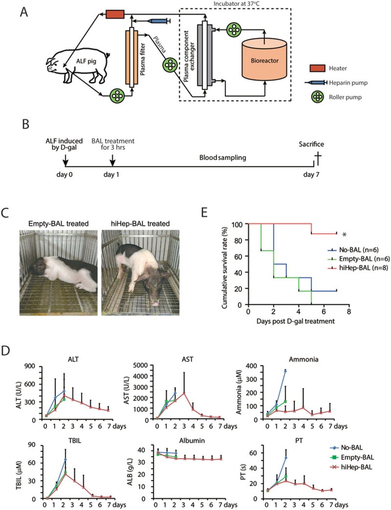
Figure 2.
hiHep-BAL rescues ALF pigs. (A) Schematic diagram depicts the configuration of the hiHep-BAL support system. Approximately 3 × 10⁹ hiHeps were implanted into the bioreactor. (B) The outline of the hiHep-BAL treatment of ALF Bama miniature pigs. (C) Bama miniature pigs treated by Empty-BAL and hiHep-BAL. Note that ALF pigs after hiHep-BAL treatment were apparently active on day 5. (D) Serum biochemical parameters of ALF pigs in No-BAL, Empty-BAL and hiHep-BAL groups. Serum levels of ALT, AST, ammonia, TBIL, albumin and prothrombin time (PT) were measured. Because most animals of No-BAL and Empty-BAL groups were dead or extremely sick from day 3, blood samples were not collected in these two groups after day 3. (E) Kaplan-Meier survival curve of No-BAL, Empty-BAL- and hiHep-BAL-treated ALF pigs (n = 6 for No-BAL and Empty-BAL, n = 8 for hiHep-BAL group). *P < 0.01, log-rank test.

