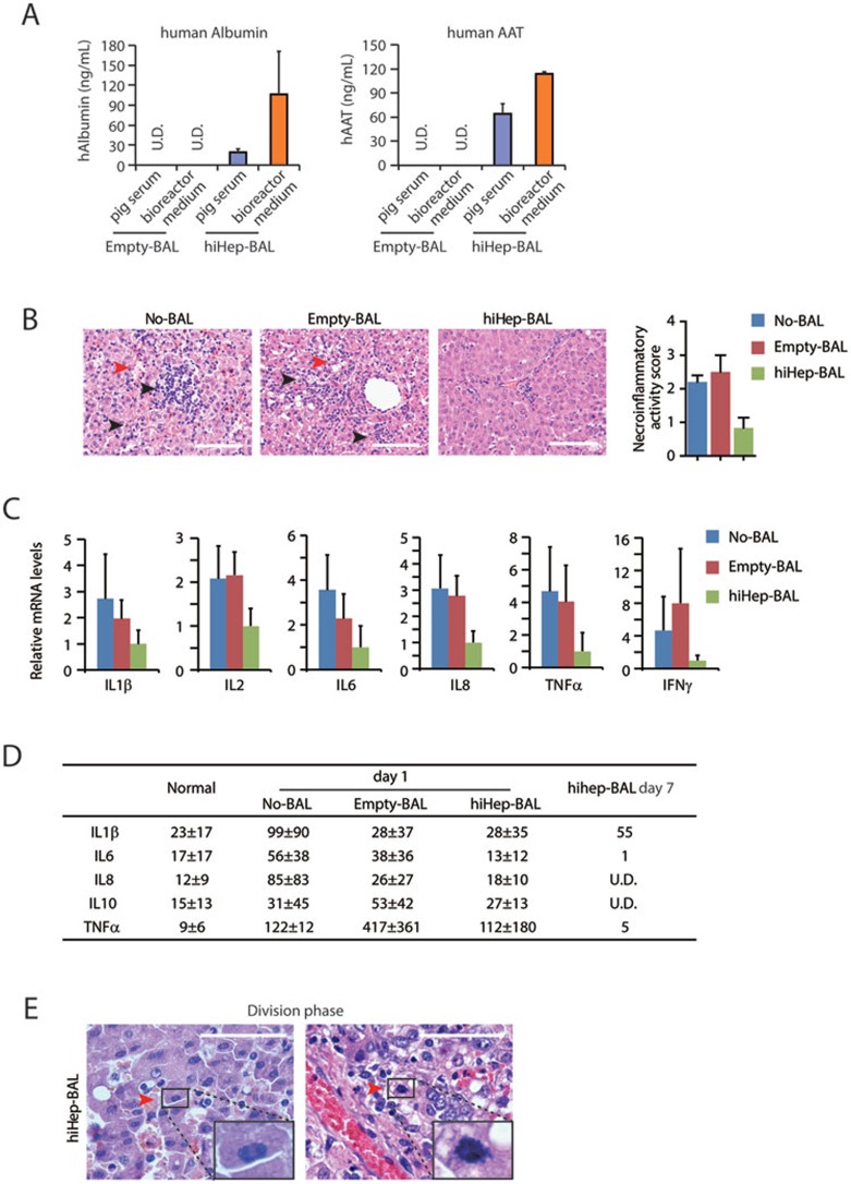
Figure 3.
Therapeutic effects of hiHep-BAL on ALF pigs. (A) Human albumin and ATT were measured in the pig serum after Empty-BAL and hiHep-BAL treatment. Human protein-specific ELISA kit was used in the assay. (B) Hematoxylin and Eosin (HE) staining of pig livers of No-BAL, Empty-BAL and hiHep-BAL groups. Livers of No-BAL and Empty-BAL groups were collected on day 2 or 3 after D-gal induction. Livers of hiHep-BAL group were collected on day 7. Red arrowheads indicate liver damage, including karyorrhexis, karyolysis and hemorrhage. Black arrowheads indicate local infiltration of inflammatory cells. Necro-inflammation was scored according to the Scheuer system. Scale bar, 100 μm. (C) mRNA expression of inflammatory cytokine genes was determined by q-PCR in pig livers of No-BAL, Empty-BAL and hiHep-BAL groups, including IL1β, IL2, IL6, IL8, TNFα and IFNγ. (D) Serum levels of the indicated cytokines were determined by ELISA with antibodies specific for pig proteins. (E) Histological analyses of proliferating hepatocytes in hiHep-BAL-treated pigs on day 7. Red arrowheads indicate hepatocytes at division phase. High magnification images of division phase are inserted.

