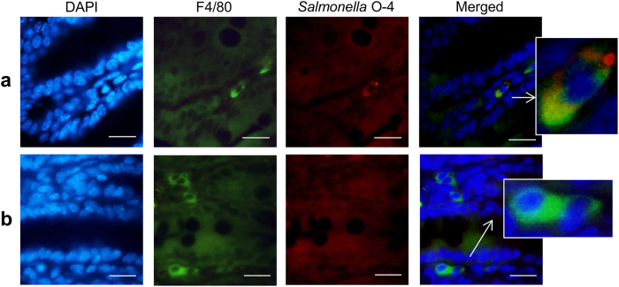
Figure 6. Salmonella colocalization with F4/80+ cells in the cecum.
Cecal sections of Salmonella-infected DBA/2J mice were stained for immunofluorescence using an F4/80 antibody targeting macrophages (green) and a Salmonella O–4 antibody (red). Nuclei were stained with DAPI (blue). Shown are representative images demonstrating (a) colocalization of Salmonella and F4/80+ cells or (b) Salmonella negative F4/80+ cells (scale bar, 20 μm).

