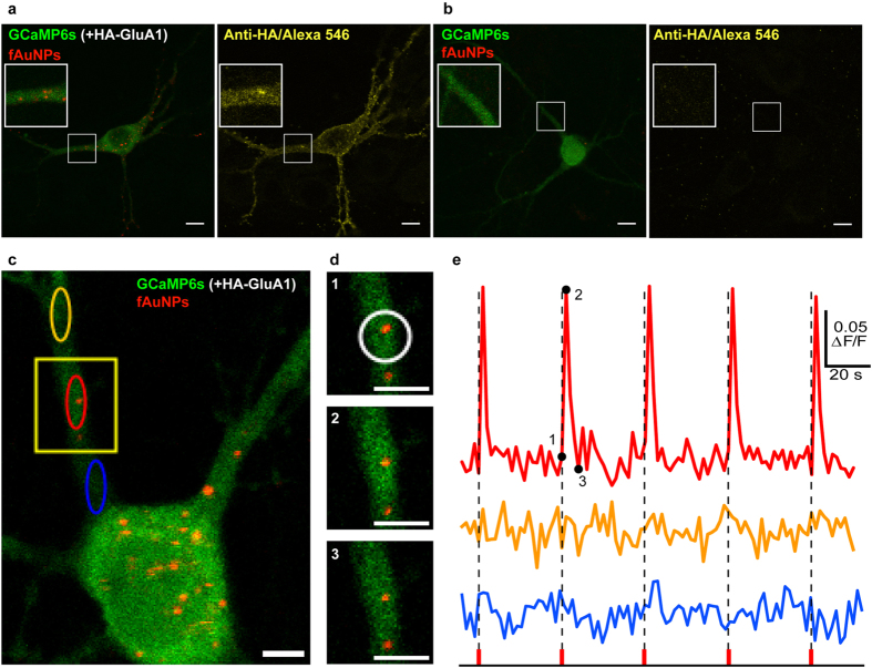
Figure 2. NALOS with functionalized AuNPs.
(a,b) Representative neurons expressing (a) HA-GluA1 and GCaMP6s (n = 5) or (b) only GCaMP6s (n = 5) and incubated 90 min with fAuNPs, imaged as in Fig. 1. The right panels in (a) and (b) show the same neuron as on their left, following fixation and immunostaining for the Ha-tag (revealed with Alexa 546). Without the presence of the HA-GluA1, the fAuNPs (functionalized with monoclonal anti-HA antibodies) did not bind on the neurons. (c-e) NALOS with fAuNPs triggered localized and repeatable Ca2+ transients (n = 10). (c) Other representative neuron transfected and imaged as in (a). (d) Magnification of the region marked in (c) for three consecutive time points (marked with 1, 2 and 3 on the graph in (e)); NALOS was applied on the region marked with a white circle. (e) Ca2+ signal (ΔF/F) of the corresponding colored oval regions marked in (c), before and after NALOS at the time points marked by dotted lines. Scale bars (a,b) 10 μm and (c,d) 5 μm.

