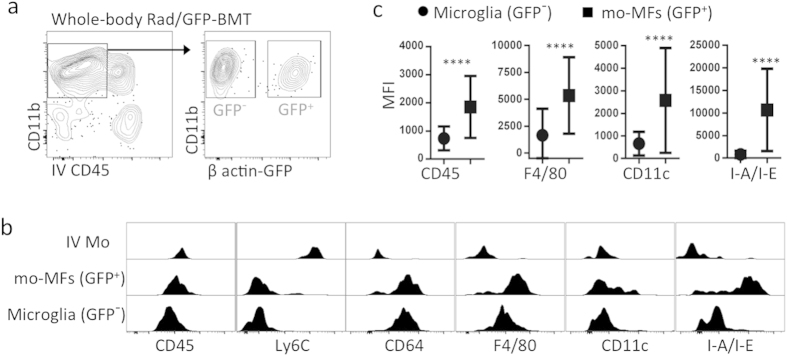
Figure 2. Microglia and recruited mo-MFs are phenotypically distinguishable in the setting of whole-body irradiation and bone marrow transplantation (Rad/BMT).
(a) Whole-body Rad/BMT leads to recruitment of myeloid cells into the retina. C57BL/6 mice were whole-body irradiated then grafted with GFP+ donor bone marrow (Rad/GFP-BMT). Using this method, we can firmly establish GFP+ cells as donor-derived whereas all GFP− cells (including microglia) are host-derived. After Rad/GFP-BMT, mice were rested for more than 3 months. Data is representative of 2 independent experiments. Cells were pre-gated on live CD45+ singlets. (b) Comprehensive phenotypic analysis reveals that microglia have a CD45lo CD11clo F4/80lo I-A/I-E− profile in the whole-body Rad/GFP-BMT setting. GFP− microglia were compared to extravascular GFP+ mo-MFs and intravascular classical Mo (IV Mo). (c) MFI indicates that the CD45lo CD11clo F4/80lo I-A/I-E− profile of GFP− microglia is significantly different from GFP+ mo-MFs. Data shown (mean ± s.d.) is a representative sample from n ≥ 4 individual samples/group. Significant differences were determined within samples by an unpaired t test (****p < 0.0001). Data is representative of 2 independent experiments.

