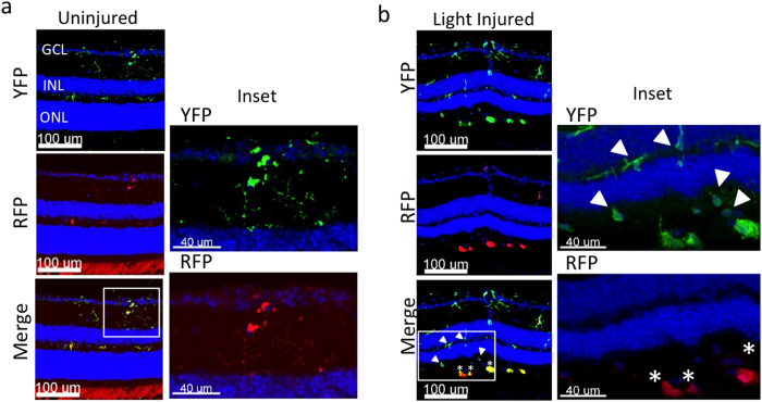
Figure 5. Fate mapping with CX3CR1YFP−CreER/wt:R26RFP mice using confocal analysis.
Following tamoxifen pulsing and a “wash out” period, mice were subjected to light challenge (or not) and retinas were harvested 5 days later. (a) Uninjured retinas contain microglia (inset: YFP+ RFP+) and display normal retinal architecture, as visualized by DAPI (outer nuclear layer = ONL; inner nuclear layer = INL; ganglion cell layer = GCL). (b) Light injured retinas have a thin ONL and possess microglia and recruited mo-MFs (inset: YFP+ RFP+ and YFP+ RFP−, respectively). Asterisks indicate microglia; arrowheads indicate recruited mo-MFs. Data is representative of two independent experiments; each experiment contained data from individual samples, uninjured n = 2 and injured n = 5.

