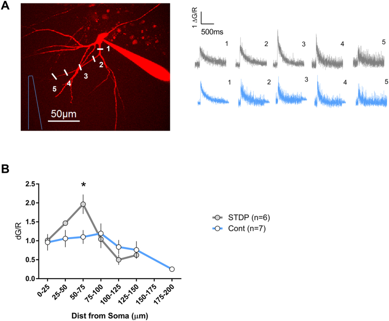
Figure 5. Local boosting of Ca2+ influx evoked by 1 backpropagating action potential in dendrites of mature granule cells after STDP conditioning protocol.
(A) Two photon fluorescence image of a mature granule cell. The inset shows representative Ca2+ transients (corresponding to the points marked in the fluorescence image in the case of the gray traces, and to similar distances for the blue traces that are corresponding to another cell in control condition) around 15–20 min after attaining the whole cell configuration. A field stimulation electrode was placed 100 μm from the soma and the closest dendrite was chosen for the measurements. (B) Profiles of backpropagating action potential-induced Ca2+ influx along dendrites of cells that received the STDP protocol (n = 6) and another group of cells with no STDP (n = 7). All recordings were made in the presence of bicuculline. *p < 0.05 Bonferroni post-hoc test after two-Way ANOVA.

